At TheHealthBoard, we're committed to delivering accurate, trustworthy information. Our expert-authored content is rigorously fact-checked and sourced from credible authorities. Discover how we uphold the highest standards in providing you with reliable knowledge.
What are Hyperintense Lesions?
Hyperintense lesions are patches of damaged cell tissue that show up as bright, white spots in certain types of specialized magnetic resonance imaging (MRI) scans. They can occur on most organs, on the brain, and along the spinal cord, and in most cases they don’t cause pain or major problems in and of themselves. They are important primarily because of how useful they are in helping to diagnose or identify the medical condition that is causing them. Multiple Sclerosis, diabetes, and dementia are among the most common, but a wide range of autoimmune and degenerative conditions could be to blame. Sometimes lesions are the first sign that something is amiss, or they could simply confirm what medical professionals already expect to see. They can’t normally be treated individually, but will often subside when the underlying problem is addressed.
Basic Presentation

In most cases hyperintense lesions are bright, glowing patches on MRI scans. They are all but invisible on the surface, so aren’t often detected during surgery, and they don’t usually show up on X-ray and computed tomography (CT) scans, either. In most cases they are quite small, often only the size of a pinprick, though radiologists are usually quick to notice them on scans thanks to their shining contrast.
How They’re Diagnosed

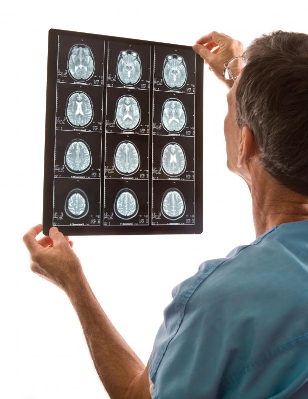
The type of MRI process that reveals these lesions is often referred to as T2-weighted MRI. MRIs use interwoven magnetic fields to create images of the all of the tissues inside a body, and are most often used to make the soft tissues appear in higher contrast than surrounding areas.
T2-weighted MR imaging uses specific settings for two factors of the imaging process: echo time and repetition time. Lesions show areas where the tissue contains more fluid than normal for the tissue type and pools of free water. It is also possible for the data from a T2 MRI to be adjusted so that the free water is not highlighted and the focus is on high concentrations of water within the tissue. This is known as a FLAIR sequence.
Common Causes

Scientists and doctors are not always sure about the exact diagnostic meaning of hyperintense lesions. They are usually a sign of some sort of larger condition, but not always. Lesions may appear many years before a larger problem actually develops in a person. Still, in most cases, they are a sign of a degenerative or autoimmune condition.
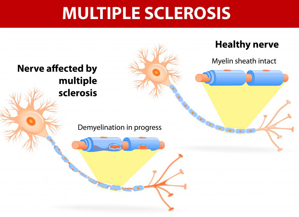
Multiple Sclerosis, a disease in which the protective coating around the body’s main nerves deteriorates, is one of the most common causes, particularly for lesions that occur along the spinal cord. Lesions in certain parts of the brain can be a sign of dementia, though this is most common in old age. Type II Diabetes and the Human Immunodeficiency Virus (HIV) and related Acquired Immunodeficiency Syndrome (AIDS) may cause spots on the liver, colon, and brain, among other places, and cancers of all forms may also be to blame.
Treatment Options

Identifying lesions is often the first step in properly diagnosing conditions, after which time doctors and healthcare providers can come up with an appropriate course of treatment. In some cases, like Creutzfeldt-Jakob disease which causes progressive dementia, the presence of hyperintense lesions can help lead to the proper diagnosis: if the lesions aren’t noticed, a person might be misdiagnosed as having typical degenerative dementia. Lesions often act as signals to care providers, helping them run the right tests and scan for the right things. Actual treatment options necessarily vary from person to person, and depend on the specifics of where the lesions are and what has likely caused them. When they’re noticed early enough, though, they can make a big difference in diagnosis and care.
AS FEATURED ON:
AS FEATURED ON:

-
![Hyperintense lesions are bright, white spots that show up on certain types of MRI scans.]() By: forestpathHyperintense lesions are bright, white spots that show up on certain types of MRI scans.
By: forestpathHyperintense lesions are bright, white spots that show up on certain types of MRI scans. -
![The radiologist or technician can view hyperintense lesions on T2 MRI images in real time from the observation room.]() By: EPSTOCKThe radiologist or technician can view hyperintense lesions on T2 MRI images in real time from the observation room.
By: EPSTOCKThe radiologist or technician can view hyperintense lesions on T2 MRI images in real time from the observation room. -
![Hyperintense lesions in certain parts of the brain can be a sign of dementia.]() By: kolotypeHyperintense lesions in certain parts of the brain can be a sign of dementia.
By: kolotypeHyperintense lesions in certain parts of the brain can be a sign of dementia. -
![Multiple sclerosis is one of the most common causes of hyperintense lesions.]() By: designuaMultiple sclerosis is one of the most common causes of hyperintense lesions.
By: designuaMultiple sclerosis is one of the most common causes of hyperintense lesions. -
![Patients who have age-related diabetes and dementia are prone to hyperintense lesions.]() By: krutoevaPatients who have age-related diabetes and dementia are prone to hyperintense lesions.
By: krutoevaPatients who have age-related diabetes and dementia are prone to hyperintense lesions.


Discussion Comments
Can anyone to explain me more about T2 hyperintense lesion up to 3.3cm in the right upper mediastinum abutting the posterolateral aspect of the superior vena cava is suspicious for an incidental mediastinal or bronchogenic cyst?
"Nonspecific T2 hyperintense lesion with a questionable thin septation within the pancreatic body measures 6 x 6 x 5 mm. Differential diagnostic considerations include an incidental epithelial cysts, side-branch IPMN, old pseudocyst, or other cystic pancreatic lesions."
What does all this mean?
I had an MRI of T-spine and signals from the liver were found. What could cause these?
what is first the memory loss or the lesion on the brain?
@MaliOhs - Hyperintense lesions on the kidney could be malignant or benign. Your doctor will have to perform more tests in order to diagnose renal cancer or other diseases.
Could a hyperintense lesion on the kidney be a sign of kidney cancer?
It is also possible that the presence of multiple hyperintense lesions on the brain can signal that a patient has secondary-progressive multiple sclerosis. More research needs to be done to determine if this is a true correlation, but if it is, it could help doctors and researchers learn more about the disease and give them another way to track the efficacy of new treatments.
Post your comments