At TheHealthBoard, we're committed to delivering accurate, trustworthy information. Our expert-authored content is rigorously fact-checked and sourced from credible authorities. Discover how we uphold the highest standards in providing you with reliable knowledge.
What Is the Brain Parenchyma?
The brain parenchyma is essentially the powerhouse of your cognitive and bodily control functions. This vital tissue comprises neurons and glial cells, which account for about 85% of the brain's weight. The remaining 15% consists of stroma, the supportive framework.
The significance of the brain parenchyma becomes evident, considering that traumatic brain injuries (TBI), which often impact the parenchyma, contribute to a substantial number of deaths and cases of permanent disability annually. Understanding the brain parenchyma and how it functions is crucial, as damage to this area can lead to profound neurological consequences.
Components

The brain parenchyma consists of neurons and glial cells. The neurons fulfill three main functions: afferent neurons are used to transmit messages from sensory organs to the brain and Central Nervous System (CNS), while efferent neurons send information and commands from the CNS to the muscles and glands. The third type, interneurons, are used for communication between the other two types.

These are supported and maintained by three types of glial cells. Oligodendroglia surround and insulate them, while astroglia physically support them and provide them with nutrition. They also eat debris and parts of dead neurons, as do microglia, the third type. Additionally, they regulate the concentration of ions in the space in between cells in the brain parenchyma, which keeps the organ as a whole functioning properly, and support the blood-brain barrier, which prevents certain substances from entering the brain via blood vessels. These cells also help with repairs following an injury.
As Compared to Stroma

The other categorization of cells in the brain is stroma, which includes blood vessels and connective tissue. It consists primarily of two sets of arteries, three sets of veins, and smaller capillaries that penetrate into the tissue, and the connective tissue that supports them. Though it doesn't perform the cognitive and management functions that the brain parenchyma does, it's still essential for the brain to function, since it provides it with nutrients and oxygen from the rest of the body. Additionally, problems with the stroma can be extremely serious. For example, if a cerebral blood vessel ruptures, the subsequent hemorrhaging can cause blood to build up in parenchymal tissue, raising the risk for stroke or memory loss.
Problems

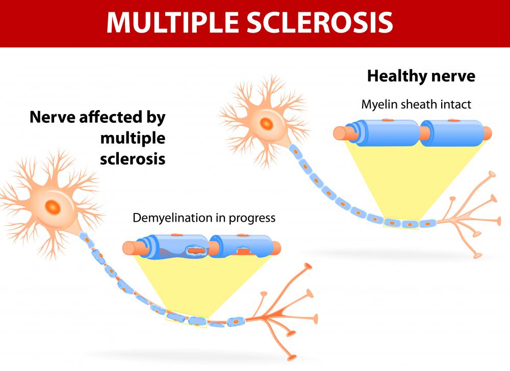
A number of different conditions can affect the brain parenchyma. Changes due to age, deterioration, trauma, or damage to the stroma can cause a wide range of conditions, including dementia, Alzheimer's, Parkinson's disease, epilepsy, multiple sclerosis, and Amyotrophic Lateral Sclerosis (ALS). Infections can also affect these cells, as in the case of encephalitis or meningitis. Additionally, cell abnormalities can lead to growths and tumors that can put pressure on or permanently damage the surrounding tissue or spread throughout the body.
What Is the Treatment of Brain Parenchyma?

Brain parenchymal treatment depends on the specific medical condition a patient faces. Generally, injuries to the brain parenchyma are known as brain lesions, which involve damaged brain tissue. There are multiple different treatments for brain lesions, including:
- Ongoing checkups to see if the lesion causes issues or grows
- Removing the brain lesion through surgery
- Using chemotherapy or radiation to treat brain tumors and cancerous lesions
- Antibiotics and other medications to treat infections
- Medication to change the immune system response
- Medicine and other treatments to alleviate symptoms of brain lesions

Central nervous system relapse including the brain parenchyma occurs in about 5 percent of patients with systemic non-Hodgkin lymphoma. The condition may also impact the spinal cord, eyes, or leptomeninges. Research shows that the best treatment for isolated central nervous relapse is systemic methotrexate.
High-dose systemic methotrexate and intrathecal chemotherapy used in combination have shown the best results.
White matter disease is also when the deepest and largest part of the brain tissue wears away. A physician may prescribe cholesterol-lowering medication or pills to help decrease your blood pressure. Also, you should not smoke when facing white matter disease.
What Is Parenchymal Brain Damage?
Parenchymal brain damage relates to the harming of functional brain tissue. Furthermore, parenchymal brain damage is a complication that takes place due to various medical conditions. It can be a serious side effect from:
- Bacterial meningitis
- Learning disabilities
- Cerebral palsy
- Mental retardation
- Seizures
- Cortical blindness
Furthermore, parenchymal brain injuries can occur among patients with abusive head trauma. Parenchymal brain damage and injury can lead to blindness, epilepsy, gross motor and cognitive impairment, and behavioral issues.
Another form of parenchymal brain damage is known as a parenchymal hemorrhage or an intraparenchymal hemorrhage, which is a bleed taking place within the functional tissues of the brain.
This happens when a blood vessel in the brain ruptures and harms the normal blood flow in the brain. As such, the patient doesn’t get an adequate amount of oxygen in brain tissues. Hypertension or high blood pressure can lead to the rupture of a blood vessel in the brain.
Cerebral amyloid angiopathy can also cause this, which is the accumulation of abnormal proteins in the brain’s arteries.
What Causes Brain Parenchymal Volume Loss?
Brain parenchymal volume loss is seen on cross-sectional imaging and is known as cerebral atrophy. Essentially, it entails losing neurons and links between those neurons. You will find various diseases can lead to brain parenchymal volume loss or brain atrophy, such as:
- Dementia
- Infection diseases
- Cerebral palsy
- Encephalitis
- HIV and AIDS
- Leukodystrophies
- Multiple sclerosis
- Stroke
- Syphilis
- Huntington’s disease and other genetic conditions
- Risk of Alzheimer’s disease
Further, head or brain injuries can lead to brain parenchymal volume loss and atrophy. Patients who smoke, drink heavily, and/or are at an advanced age tend to increase their risk of brain atrophy. In addition, brain atrophy can lead to dementia among older patients.
They will struggle with thinking and remembering to the point of finding everyday life overly difficult. Such patients can get Alzheimer’s disease due to brain atrophy. These neurodegenerative diseases may cause aphasia, which involves speaking and language issues.
Brain atrophy can also lead to seizures, loss of consciousness, and bitter or metallic tastes.
What Is a Parenchymal Stroke?
A parenchymal stroke is a type of hemorrhagic stroke. The two other types of hemorrhagic strokes include arteriovenous and subarachnoid malformations. A parenchymal hemorrhage is a brain bleed that takes place within the parenchymal tissues. It can disrupt blood oxygen flow in brain cells and even lead to the destruction of functional brain tissue.
An intraparenchymal hemorrhage is responsible for less than 20 percent of all strokes, but it does have the highest mortality risks of all stroke types. Headaches, seizures, and neurological deficits can occur from a parenchymal stroke. Patients often have speech, vision, and hearing issues.
The causes of hemorrhagic strokes include smoking, drinking alcohol heavily, and having high blood pressure or hypertension. Doctors can conduct blood coagulation studies and imaging studies to determine if a patient has a parenchymal hemorrhage.
Physicians may prescribe blood-thinning medication or anticoagulants along with mannitol or hypertonic saline. Platelet therapy is another option for treating that type of stroke.
FAQ on Brain Parenchyma
What is the brain parenchyma?
The brain parenchyma refers to the functional tissue in the brain that is made up of two main types of cells: neurons and glial cells. Neurons are responsible for transmitting information through electrical and chemical signals, while glial cells provide support and protection for neurons. The brain parenchyma is critical for all brain activities, including thought, memory, emotion, and coordination.
How does the brain parenchyma differ from other brain structures?
Unlike other brain structures such as the meninges (protective layers) or blood vessels, the brain parenchyma specifically denotes the brain's functional tissue. It is the core substance within the central nervous system where the processing of information occurs. Other structures serve supportive or protective roles, but the parenchyma is where the actual 'work' of the brain takes place.
Can the brain parenchyma be damaged, and what are the consequences?
Yes, the brain parenchyma can be damaged due to various causes such as trauma, stroke, infection, or degenerative diseases. Damage to the parenchyma can lead to a loss of function in the affected area, which may manifest as cognitive deficits, sensory loss, or motor dysfunction, depending on the location and extent of the damage. According to the Centers for Disease Control and Prevention (CDC), stroke, a common cause of brain parenchymal damage, affects approximately 795,000 people in the United States each year.
What imaging techniques are used to assess the brain parenchyma?
Imaging techniques such as Magnetic Resonance Imaging (MRI) and Computed Tomography (CT) scans are commonly used to assess the brain parenchyma. MRI provides high-resolution images and can detect changes in brain tissue, such as inflammation, bleeding, or scarring. CT scans are faster and can quickly identify fractures or bleeding. Advanced imaging techniques like Diffusion Tensor Imaging (DTI) can even map neural pathways, providing detailed insights into brain structure and function.
How does aging affect the brain parenchyma?
Aging can lead to changes in the brain parenchyma, including a reduction in brain volume and weight. This is partly due to the loss of neurons and the shrinking of neuron size. There may also be a decrease in the number of synaptic connections, which can affect cognitive functions. However, the brain can adapt to some of these changes through neuroplasticity, which allows it to reorganize and form new connections. According to a study published in the journal Nature Reviews Neuroscience, the human brain experiences a roughly 5% decrease in volume per decade after the age of 40.
AS FEATURED ON:
AS FEATURED ON:

-
![Changes in age can affect the way tissues function in the brain, also known as brain parenchyma.]() By: ArtanikaChanges in age can affect the way tissues function in the brain, also known as brain parenchyma.
By: ArtanikaChanges in age can affect the way tissues function in the brain, also known as brain parenchyma. -
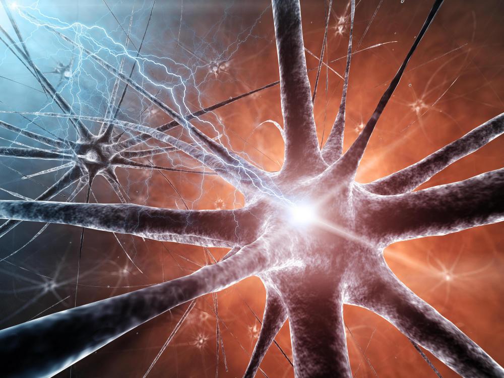
![Brain parenchyma is partially made up of neurons that communicate with organs or muscles of the body.]() By: nobeastsofierceBrain parenchyma is partially made up of neurons that communicate with organs or muscles of the body.
By: nobeastsofierceBrain parenchyma is partially made up of neurons that communicate with organs or muscles of the body. -
![Functional brain tissue has different cells that control cognition and body functions.]() By: shutswisFunctional brain tissue has different cells that control cognition and body functions.
By: shutswisFunctional brain tissue has different cells that control cognition and body functions. -
![Trauma to the brain parenchyma can result in decreased cognitive functions or even death.]() By: WavebreakmediaMicroTrauma to the brain parenchyma can result in decreased cognitive functions or even death.
By: WavebreakmediaMicroTrauma to the brain parenchyma can result in decreased cognitive functions or even death. -
![Damage to the brain parenchyma often results in a loss of cognitive ability or even death.]() By: Verity JohnsonDamage to the brain parenchyma often results in a loss of cognitive ability or even death.
By: Verity JohnsonDamage to the brain parenchyma often results in a loss of cognitive ability or even death. -
![Diseases like multiple sclerosis can affect brain parenchyma.]() By: designuaDiseases like multiple sclerosis can affect brain parenchyma.
By: designuaDiseases like multiple sclerosis can affect brain parenchyma.


Discussion Comments
@MrsPramm: I would tend to agree with you. In fact, it is very easy to prove. We only need to answer the question 'how does a thought originate?' or 'where does the SA node in our heart get the energy to generate the first pulse of a heart cycle at almost clock like precision?'
My view is the brain perceives and paints a picture for the soul to act. In dementia or Alzheimer's, the ability of brain to paint this picture is diminished/lost. And the soul (being energy itself) provides the energy to the SA node.
@umbra21 - Well, it's a good thing to remember that the brain isn't magical. Often when people gain what seems like amazing abilities from brain damage, what they've actually gained is an intense focus on a particular skill, sometimes to the detriment of other things, and that leads them to become extraordinary at that skill. An ordinary person could achieve the same thing if they were similarly focused.
The brain is still somewhat mysterious, but we actually know a huge amount about it now. There is a lot of misinformation about it around though.
@MrsPramm - I'm sure some people would debate that everything we consider to be a soul is contained in the brain. I would prefer if it wasn't true myself. When you think about how vulnerable the brain is to disease and damage, it makes you kind of question who a person actually is deep inside.
Is an Alzheimer patient losing themselves, or is it only that they have a kind of disability and their soul is still intact? I like to think that they remain who they are and the brain is only the trappings of that.
But it is definitely fascinating reading about people who have gained or lost abilities because of brain damage, particularly when they gain abilities. It makes you wonder what talents are inherent in all of us, hidden in our brains, in the mass of grey matter.
It's incredible how many different facets there are to the human brain. I mean, you've got the physical aspects to it here, the gross structures and the cells and how they are all connected to each other and to the rest of the body.
And then you've got the way they work, and how we experience it. When you think about how everything that a human would recognize as being a part of soul or consciousness is contained in the cells that make up the brain parenchyma. And they are just a sort of formless looking (to the layman, anyway) grey mass, but are also so very special.
Post your comments